Are Dental X-Rays Safe? | EndoCare Dental Clinic, Viman Nagar Pune
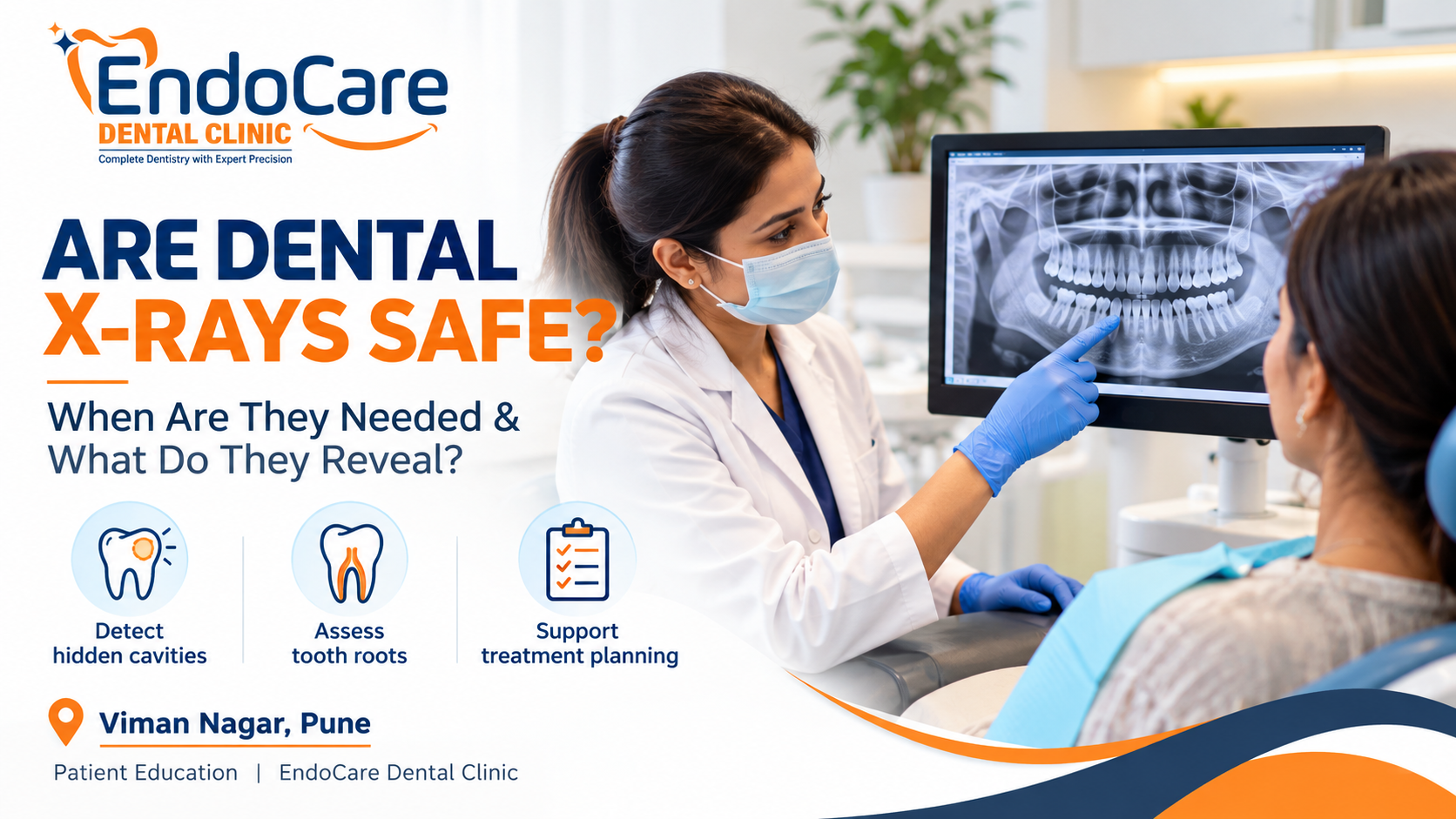
Dental X-Rays in Viman Nagar, Pune: When Are They Needed and Are They Safe?
Have you ever visited a dentist for a routine check-up and been advised to get a dental X-ray even though your teeth looked perfectly fine?
It is a common question among patients: Why do I need a dental X-ray if my dentist can already examine my teeth?
The answer is that some dental problems develop in places that cannot be assessed fully by looking inside the mouth alone. Cavities between teeth, changes around tooth roots, certain infections and problems involving the supporting bone may require additional imaging to evaluate properly.
However, dental X-rays are not necessary at every appointment. Whether you need one depends on your symptoms, dental history, examination findings and individual risk of oral disease.
At EndoCare Dental Clinic in Viman Nagar, Pune, understanding the purpose of recommended investigations can help patients make informed decisions about their dental care.
This guide explains what dental X-rays can reveal, when dentists recommend them, the different types of dental imaging, and what patients should know about radiation safety.
In This Article
- Why might a dentist recommend dental X-rays?
- What can dental X-rays reveal?
- What are the different types of dental X-rays?
- Are dental X-rays safe?
- Are dental X-rays safe during pregnancy?
- Do children need dental X-rays?
- How often should you get dental X-rays?
- What should you tell your dentist before an X-ray?
- Frequently asked questions
Why Might a Dentist Recommend Dental X-Rays?
A dental examination allows your dentist to assess visible teeth, gums, existing restorations and other features of your oral health.
However, a visual examination does not always reveal everything happening beneath the tooth surface or around the roots.
Your dentist may recommend dental X-rays when the additional information could help establish a diagnosis, assess a problem or plan treatment.
Common reasons include:
- Suspected cavities between teeth: Early decay between neighbouring teeth may be difficult to identify through visual examination alone.
- Persistent tooth sensitivity or pain: Imaging may help assess the roots and surrounding tissues when a problem is suspected.
- Possible infection around a tooth root: An X-ray may reveal changes in the bone surrounding the root, although the findings must be interpreted alongside the clinical examination.
- Assessment before root canal treatment: Imaging can help the dentist assess root anatomy and changes around the affected tooth.
- Gum and bone assessment: Certain radiographs can help evaluate the supporting bone around teeth.
- Impacted or unerupted teeth: Imaging may help determine the position of teeth that have not erupted normally.
- Dental injury: A dentist may recommend imaging when a tooth or surrounding structures have been injured.
- Treatment planning: Certain restorative, surgical or implant-related procedures may require imaging appropriate to the clinical question.
Not every patient with these concerns needs the same type of X-ray. Your dentist should explain why a particular image is being recommended and how it may influence your treatment.
What Can Dental X-Rays Reveal?
Dental radiographs provide information that may not be visible during an ordinary mouth examination.
1. Cavities Between Teeth
Dental decay can develop on surfaces where two teeth touch. These areas may be difficult to inspect directly, particularly when the contact between the teeth is tight.
Depending on the location and clinical findings, bitewing radiographs can help identify decay between teeth and assess its extent.
Detecting a cavity does not automatically mean that a filling is required. Your dentist will consider the appearance of the lesion, its location, progression risk and other examination findings when deciding what to do.
2. Problems Around Tooth Roots
The roots of your teeth extend into the jawbone and cannot be examined completely by looking at the visible crown.
When a dentist suspects inflammation or infection involving the tissues around a tooth root, an appropriate radiograph may help identify changes in the surrounding bone.
This information can contribute to decisions about whether a tooth needs monitoring, restorative treatment or further investigation.
3. Bone Changes Associated With Gum Disease
Certain dental X-rays can show the level and pattern of the bone supporting the teeth.
These findings can help a dentist assess the extent of periodontal damage when considered alongside gum measurements, bleeding, tooth mobility and other clinical signs.
An X-ray alone cannot fully determine the health of your gums.
4. Impacted Teeth and Tooth Position
Some teeth do not erupt into their expected positions. Wisdom teeth are a familiar example.
Imaging may help establish the position of an impacted tooth and its relationship to neighbouring teeth and surrounding structures.
The type of image required depends on the clinical question. Three-dimensional imaging is not automatically necessary for every impacted tooth.
5. Planning Dental Treatment
Dental imaging can also assist with treatment planning.
For example, a dentist may use appropriate radiographs when assessing a tooth before root canal treatment or determining whether additional imaging is needed for a more complex procedure.
The objective is not to take as many images as possible. It is to obtain the information needed to make a sound clinical decision.
What Are the Different Types of Dental X-Rays?
Different radiographs show different parts of the teeth and jaws. The dentist chooses the type according to the information needed.
1. Bitewing X-Rays
Bitewing images usually show the crowns of the upper and lower back teeth in the same region.
They are commonly used to assess cavities between teeth and can also help evaluate supporting bone levels.
Your dentist may recommend bitewings when these areas cannot be assessed adequately through a visual examination alone.
2. Periapical X-Rays
A periapical radiograph shows a tooth from its visible crown to the tip of its root, together with some surrounding bone.
It may be useful when investigating pain, suspected root-related disease, dental trauma or findings associated with root canal treatment.
The dentist selects the area to be imaged according to the symptoms and examination.
3. Panoramic Dental X-Rays
A panoramic radiograph provides a broad image of the teeth, jaws and surrounding structures.
It may be recommended for selected assessments involving impacted teeth, jaw structures or broader treatment planning.
However, a panoramic image does not provide the same detailed view of every tooth surface as a targeted intraoral radiograph. It is not necessarily the best choice for detecting every small cavity.
4. Cone-Beam Computed Tomography (CBCT)
CBCT is a specialised form of dental imaging that produces three-dimensional views of selected oral and maxillofacial structures.
It may be considered in certain complex situations, such as selected implant-planning cases or when conventional two-dimensional images do not provide enough information to answer an important clinical question.
CBCT generally exposes patients to more radiation than conventional dental X-rays. It should therefore be used when the additional three-dimensional information is clinically justified, not as a routine replacement for ordinary dental radiographs. U.S. Food and Drug Administration
The important distinction: A more advanced scan is not automatically a better scan for every patient. The appropriate investigation is the one that answers the clinical question with the least unnecessary exposure.
Are Dental X-Rays Safe?
This is one of the most common concerns patients raise before dental imaging.
Dental X-rays use ionising radiation, so the exposure is not literally zero-risk. However, conventional dental radiographs generally involve low radiation doses, and the clinical benefit can outweigh the small potential risk when imaging is appropriately indicated.
The American Dental Association and the US Food and Drug Administration recommend selecting dental radiographs according to individual clinical needs and minimising unnecessary exposure. U.S. Food and Drug Administration
There are three important principles to understand.
1. Dental X-Rays Should Have a Clear Clinical Purpose
Your dentist should consider your symptoms, examination findings, dental history and risk of disease before recommending an X-ray.
A radiograph should be taken when the additional information is expected to contribute meaningfully to your care.
Routine imaging without an appropriate clinical reason should be avoided.
2. Radiation Exposure Should Be Minimized
Dental professionals should follow appropriate radiation-protection practices and use the minimum exposure necessary to obtain a diagnostically useful image.
Digital imaging can offer advantages, including the potential for lower radiation exposure compared with conventional film systems, depending on the equipment and technique used. However, digital technology does not eliminate radiation exposure or justify unnecessary images. U.S. Food and Drug Administration
3. Previous X-Rays May Be Relevant
If you have recently had dental X-rays, tell your dentist and bring previous images or records if available.
Your dentist can assess whether the earlier images are suitable for the current clinical question or whether new imaging is necessary.
Avoiding unnecessary duplicate images is one practical way to reduce avoidable exposure.
Should You Refuse Dental X-Rays Because of Radiation?
Not automatically.
Declining a clinically important investigation can leave a dental problem incompletely assessed. At the same time, taking an X-ray simply because it is customary, without considering your individual needs, is not appropriate either.
If you are worried about radiation, ask your dentist:
- What are you looking for in this X-ray?
- How will the result affect my treatment?
- Is an existing image sufficient?
- Is this the most appropriate type of imaging for my situation?
A clear explanation should help you understand the benefits, limitations and reasons for the recommendation.
Are Dental X-Rays Safe During Pregnancy?
Pregnancy is an important consideration to discuss with your dentist before any dental imaging.
If you are pregnant or think you may be pregnant, tell your dentist before the examination. This allows the clinician to consider your circumstances, the urgency of the dental problem and whether the proposed imaging is justified.
Pregnancy does not mean that every dental X-ray must automatically be postponed. Necessary dental investigations may still be appropriate when they are expected to affect diagnosis or treatment.
The decision should be made by your treating dental professional, taking account of the clinical need and applicable radiation-protection guidance.
Do not ignore severe tooth pain, facial swelling or other concerning symptoms simply because you are pregnant. Contact a dental professional for advice about the safest way to assess and manage the problem.
Do Children Need Dental X-Rays?
Children do not automatically need dental X-rays at every dental appointment.
A dentist may recommend imaging when it is needed to assess a particular concern, such as:
- Cavities that cannot be evaluated adequately by visual examination.
- The development or position of teeth.
- An injury involving a tooth.
- Suspected dental disease that requires further investigation.
- A specific treatment-planning question.
The decision depends on the child's age, stage of dental development, examination findings and risk of disease.
Radiographs should be justified individually, and exposure settings should be appropriate for the child and the imaging task. The FDA advises particular care to minimise unnecessary radiation exposure in children. U.S. Food and Drug Administration
Parents can help by sharing previous dental images and asking why the proposed radiograph is needed.
How Often Should You Get Dental X-Rays?
There is no single schedule that is appropriate for every patient.
Some people may need imaging because of symptoms or an increased risk of dental disease. Others with stable oral health may not need new radiographs at a particular routine visit.
Your dentist considers factors such as:
- Your current dental health.
- Previous dental problems and treatment.
- Your risk of developing cavities.
- Symptoms such as pain or sensitivity.
- Whether a previous image is still useful.
- The specific information required for diagnosis or treatment.
For this reason, dental X-rays should not automatically be repeated at every six-month check-up.
The ADA's guidance emphasises individual assessment rather than a universal interval for all patients. American Dental Association
A useful rule: Ask whether a new image is needed for your current dental condition, rather than assuming that every check-up requires one.
What Should You Tell Your Dentist Before an X-Ray?
Before dental imaging, share any information that may help your dentist make an appropriate decision.
Tell your dentist if:
- You are pregnant or think you may be pregnant.
- You have had recent dental X-rays or other relevant imaging.
- You are experiencing pain, swelling, sensitivity or a recent dental injury.
- You have undergone previous treatment on the affected tooth.
- You have questions about why the image is being recommended.
If you have previous images, bring them along when possible. The dentist can decide whether they provide sufficient information or whether new imaging is needed.
You can also ask what the radiograph is expected to show and how its findings may change the treatment plan.
Understanding the purpose of the investigation can make the dental visit more reassuring and help you participate in decisions about your care.
Dental X-Rays in Viman Nagar, Pune: What Should Patients Expect?
If you are visiting a dental clinic in Viman Nagar, Pune, you can ask your dentist to explain any recommended X-ray before it is taken.
A useful discussion should cover the reason for imaging, the type of radiograph proposed and how the findings will contribute to diagnosis or treatment planning.
At EndoCare Dental Clinic, dental assessment may involve reviewing symptoms, examining the affected area and considering whether appropriate imaging is needed.
For example, if a dentist suspects a cavity, an X-ray may help assess areas that are difficult to inspect visually. If root canal treatment is being considered, imaging may help evaluate the affected tooth and surrounding structures.
The exact investigation depends on the individual case. Not every patient requires the same images, and more extensive imaging is not automatically necessary.
EndoCare Dental Clinic is located in Viman Nagar, Pune, and serves patients from nearby areas including Kalyani Nagar, Wadgaon Sheri, Kharadi and Dhanori.
If you have questions about a recommended dental X-ray, ask your dentist to explain its purpose and how the findings will influence your care.
Frequently Asked Questions About Dental X-Rays
1. Are dental X-rays safe for adults?
Conventional dental X-rays generally involve low radiation exposure. They should be recommended when clinically justified, with appropriate precautions to minimise unnecessary exposure. Your dentist can explain why imaging is appropriate for your particular situation.
2. Can dental X-rays detect cavities that a dentist cannot see?
Yes. Certain radiographs, particularly bitewing images, can help detect cavities between teeth that may not be visible during an ordinary examination. However, the need for imaging depends on your individual clinical findings.
3. Do I need dental X-rays at every check-up?
No. The decision depends on your oral health, symptoms, previous images and risk of dental disease. There is no universal requirement to take new X-rays at every routine visit.
4. Are dental X-rays safe during pregnancy?
Dental imaging may be appropriate during pregnancy when it is clinically necessary. Tell your dentist that you are pregnant or may be pregnant so that the need for imaging and appropriate precautions can be assessed.
5. Are dental X-rays necessary before root canal treatment?
Dental radiographs are commonly used to help assess a tooth before root canal treatment. They can provide information about the roots and surrounding tissues. The dentist determines the type and extent of imaging required for the individual case.
6. What is the difference between a dental X-ray and a CBCT scan?
Conventional dental X-rays usually provide two-dimensional images. CBCT produces three-dimensional images and may be useful for selected complex diagnostic or treatment-planning questions. CBCT generally involves more radiation than conventional dental X-rays, so it should be used only when the additional information is justified. U.S. Food and Drug Administration
Make Informed Decisions About Your Dental Health
Dental X-rays can help dentists identify problems that may not be visible during an ordinary examination. They can contribute to the assessment of cavities, tooth roots, supporting bone and selected treatment-planning questions.
However, the right approach is not to take more X-rays, but to take the images that are genuinely needed for your care.
If your dentist recommends an X-ray, ask why it is needed, what information it is expected to provide and how the result may influence your treatment.
If you are looking for a dental consultation in Viman Nagar, Pune, EndoCare Dental Clinic can help you discuss your dental concerns and understand the assessment and treatment options appropriate to your condition.
EndoCare Dental Clinic, Viman Nagar Pune
- Phone: +91 90968 82602
- Hours: Open all seven days, 10:00 AM–9:00 PM
- Appointment: Contact the clinic to arrange a consultation.
You can also visit the clinic's Contact and Appointments page for appointment information.
Medical disclaimer: This article is intended for general educational purposes and does not replace an individual dental examination, diagnosis or treatment recommendation. Your dentist should determine whether dental imaging is appropriate for your circumstances.
This article is general information from the EndoCare Dental Clinic team. It is not a diagnosis or a substitute for an examination by your dentist. Please consult a dentist for advice about your own teeth.